Chest wall disorders can significantly affect respiratory function, leading to a variety of challenges in daily life. These conditions often stem from irregularities in the chest wall structure, which can be congenital or acquired over time. Understanding how these abnormalities impact breathing is crucial for both patients and healthcare providers.
From the distinctive sunken appearance of pectus excavatum to the prominence of pectus carinatum, each disorder presents unique symptoms and treatment options. As we delve into this topic, we will explore various types of chest wall disorders, their causes, and what management strategies are available to improve quality of life for those affected. Whether you’re a patient seeking information, or someone interested in medical insights, this comprehensive guide will shed light on an area that deserves attention!
Understanding Chest Wall Disorders: An Overview
Chest wall disorders encompass a range of conditions that affect the structure and function of the chest. These abnormalities can hinder respiratory efficiency, leading to complications in breathing and overall health.
The chest wall consists of bones, muscles, and cartilage that work together to protect vital organs like the heart and lungs. When this intricate system is disrupted, patients may experience various symptoms beyond just physical deformities.
Causes vary widely; some disorders are present at birth while others develop over time due to factors such as injury or obesity. Recognizing these issues early on is essential for effective management.
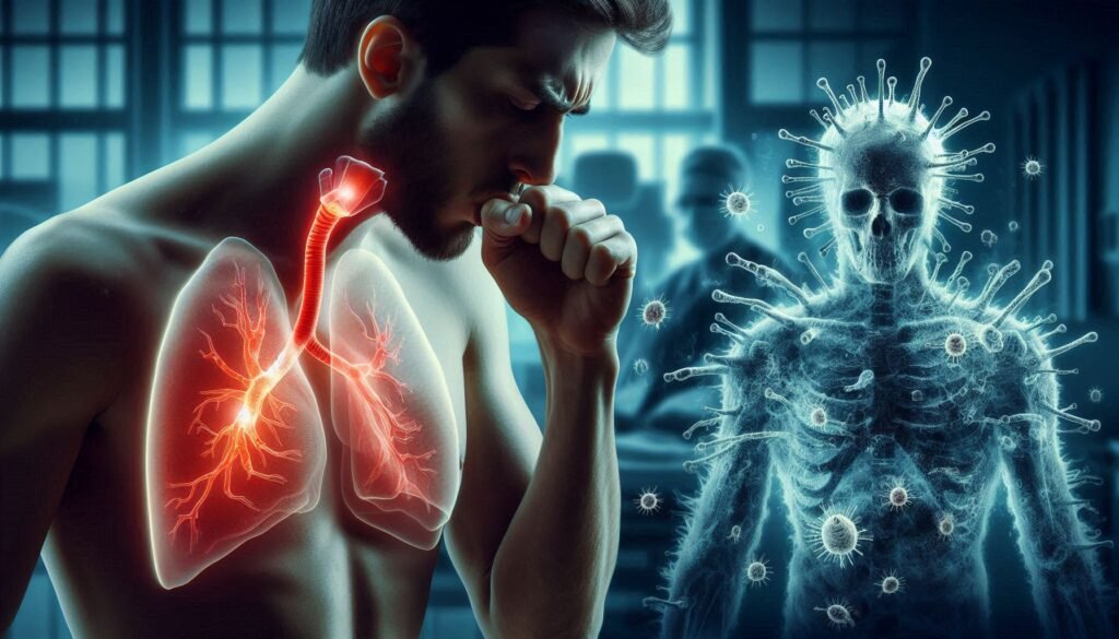
“Why Does Atelectasis Cause Breathing Problems? Understanding Lung Collapse”
The impact extends beyond respiratory limitations. Chest wall disorders can influence self-esteem, social interactions, and emotional well-being. Understanding these complexities paves the way for better awareness and treatment options tailored to individual needs.
Anatomy of the Chest Wall: Structure and Function
The chest wall is a complex structure essential for respiratory function. It comprises bones, muscles, and tissues that protect vital organs like the heart and lungs.
The rib cage forms the bony framework. Twelve pairs of ribs attach to the spine at the back and curve around to connect with the sternum in front. This arrangement provides stability while allowing flexibility for breathing.
“What Makes Pulmonary Fibrosis Progressive? Treatment & Management”
Intercostal muscles sit between each rib pair. They play a critical role during inhalation and exhalation by expanding and contracting the thoracic cavity. The diaphragm, located beneath these structures, acts as a primary muscle responsible for drawing air into the lungs.
In addition to its protective features, the chest wall helps regulate pressure changes necessary for effective ventilation. Any irregularities in this anatomy can significantly impact respiratory health and overall well-being.
Types of Chest Wall Disorders: From Congenital to Acquired
Chest wall disorders can be categorized into two main types: congenital and acquired. Congenital disorders are present at birth, stemming from developmental anomalies during gestation. Examples include pectus excavatum and pectus carinatum, where structural deformities of the rib cage affect its shape and function.
“How Does Myasthenia Gravis Affect Your Breathing? Key Information”
Acquired chest wall disorders result from various external factors or conditions that develop over time. These may arise due to trauma, such as flail chest following an accident, or progressive diseases like kyphoscoliosis, which involves abnormal spine curvature impacting respiratory capacity.
Other acquired issues can stem from obesity, leading to excess weight that restricts lung expansion. Each disorder presents unique challenges but shares a commonality in potentially compromising respiratory function. Prompt diagnosis and tailored management strategies are essential for optimal outcomes in individuals affected by these conditions.
Pectus Excavatum: Causes, Symptoms, and Treatment Options
Pectus excavatum, often referred to as “sunken chest,” is a common congenital deformity. It occurs when the breastbone sinks into the chest cavity. The exact cause remains unclear, but genetics play a significant role.
“What Causes Sudden Dyspnea? Understanding Breathing Difficulties”
Symptoms can vary greatly among individuals. Some may experience no issues at all, while others might face respiratory problems or cardiovascular complications due to restricted lung capacity. Physical appearance can also lead to psychological impacts, particularly in adolescents.
Treatment options depend on severity and symptoms. Mild cases may only require monitoring, while more severe instances often warrant surgical intervention. The Nuss procedure involves placing a bar under the ribcage to lift it into place, whereas the Ravitch technique removes abnormal cartilage for correction.
Non-surgical treatments like braces are available for younger patients still growing. Each case requires careful assessment by healthcare professionals to determine the best approach tailored to individual needs.
Pectus Carinatum: Recognizing and Managing the “Pigeon Chest”
Pectus carinatum, often referred to as “pigeon chest,” is a condition characterized by an abnormal protrusion of the sternum. This deformity can be noticeable during childhood or early adolescence and may lead to both physical and psychological challenges.
“Why Is My Breathing So Fast? Understanding Tachypnea”
The exact cause remains unclear, but genetic factors are believed to play a role. Some children with pectus carinatum experience no symptoms, while others may face discomfort or issues with their self-esteem due to the visible changes in their chest shape.
Management options vary based on severity. Mild cases might only require monitoring, while more pronounced deformities could benefit from bracing techniques designed to gradually correct the protrusion over time. In severe instances where breathing or cardiac function is compromised, surgical intervention may become necessary.
“What Are the Dangers of Hypoxemia? Signs You Need Medical Help”
Supportive therapies such as counseling can also help address emotional concerns associated with this condition. Early recognition and intervention pave the way for better outcomes in affected individuals.
Kyphoscoliosis: When Spine Curvature Affects Breathing
Kyphoscoliosis is a complex spinal deformity characterized by an abnormal curvature of the spine. It combines both kyphosis, which is an exaggerated forward rounding of the back, and scoliosis, a sideways curvature. This dual condition can significantly impact respiratory function.
As the spine curves abnormally, it compresses thoracic cavity space. This leads to restricted lung capacity and impaired breathing mechanics. Patients may experience shortness of breath or fatigue during physical activity.
In severe cases, kyphoscoliosis may result in structural changes that affect vital organs beyond just the lungs. Regular monitoring becomes crucial for managing symptoms and preventing complications over time.
“What Causes Altitude Sickness? Prevention & Treatment Guide”
Treatment options vary based on severity but often include physical therapy aimed at strengthening core muscles. In some instances, surgical intervention may be necessary to correct spinal alignment and improve respiratory efficiency.
Flail Chest: Causes, Diagnosis, and Emergency Management
Flail chest is a serious condition resulting from multiple rib fractures. It typically occurs due to blunt trauma, such as in car accidents or falls. This injury leads to a segment of the chest wall becoming detached, causing paradoxical movement during breathing.
“Is Epiglottitis Affecting Your Breathing? Emergency Warning Signs”
Diagnosis begins with physical examination and imaging techniques like X-rays or CT scans. Doctors look for rib deformity and assess lung function to gauge the severity of the injury. Symptoms include severe pain, difficulty breathing, and sometimes visible chest wall movement that contradicts normal respiration.
Emergency management focuses on stabilizing the patient’s condition. Providing supplemental oxygen is crucial if respiratory distress is evident. In some cases, mechanical ventilation may be necessary until adequate healing occurs. Pain control through medications helps improve comfort and facilitate deeper breaths, which are essential for recovery and preventing further complications.
Thoracoplasty: Understanding Surgical Chest Wall Modifications
Thoracoplasty is a surgical procedure aimed at correcting chest wall deformities. By removing or reshaping portions of the rib cage, surgeons can enhance both functionality and appearance.
“What Makes Pulmonary Edema Life-Threatening? Critical Signs”
This intervention often addresses severe conditions like pectus excavatum or flail chest, where structural abnormalities compromise respiratory function. The surgery may involve ribs being resectioned to allow for better lung expansion.
Recovery typically involves pain management and physical therapy to restore movement in the thoracic region. Patients often experience improved breathing capacity following healing.
Surgeons use advanced imaging techniques preoperatively to plan the best approach tailored to each individual’s needs. This precision helps minimize complications and optimize outcomes.
Patients should discuss potential risks with their healthcare provider, as every surgery carries inherent uncertainties. Thoracoplasty not only aims for aesthetic improvements but also plays a crucial role in enhancing quality of life by stabilizing the chest wall structure.
The Impact of Obesity on Chest Wall Function
Obesity can significantly impact chest wall function, altering respiratory mechanics. Excess fat accumulation around the thoracic cavity restricts lung expansion. This limitation can lead to decreased lung volumes.
“How Does Emphysema Progress? Understanding Lung Damage”
The added weight increases the work of breathing. Patients may experience shortness of breath during minimal activity due to this burden. As a result, their overall exercise tolerance often declines.
Moreover, obesity predisposes individuals to obstructive sleep apnea, exacerbating breathing difficulties at night. The combination of elevated body mass and compromised airways creates a cycle that further impairs pulmonary function.
These changes can increase the risk for various cardiovascular conditions as well. Maintaining a healthy weight is essential not only for general well-being but also for optimal respiratory health in those affected by obesity-related issues with their chest walls.
Diagnostic Approaches: Imaging Techniques and Pulmonary Function Tests
Diagnosing chest wall disorders involves a careful selection of imaging techniques and pulmonary function tests. These methods provide crucial insights into structural abnormalities.
X-rays are typically the first line of evaluation. They reveal bone structure and can highlight deformities like pectus excavatum or carinatum. However, for more detailed assessments, CT scans are preferred. They offer cross-sectional images that allow physicians to evaluate both soft tissues and bony structures.
Pulmonary function tests (PFTs) complement imaging by assessing lung capacity and airflow. These tests measure how well your lungs work, helping to identify any respiratory limitations caused by chest wall deformities.
Together, these diagnostic approaches enable healthcare providers to formulate effective treatment plans tailored to each patient’s needs. By understanding the specific nature of the disorder, clinicians can address functional impairments efficiently.
Surgical Interventions: When and How to Correct Chest Wall Abnormalities
Surgical interventions for chest wall disorders are crucial when conservative treatments fail to improve respiratory function or quality of life. The decision to pursue surgery often hinges on the severity of the deformity and its impact on lung capacity.
One common procedure is the Nuss procedure, primarily used for pectus excavatum. This minimally invasive approach involves placing a curved metal bar beneath the sternum to elevate it into a more normal position.
For conditions like pectus carinatum, surgeons may perform an open surgical repair called resection and sternal flattening. This technique corrects protruding structures effectively while minimizing recurrence rates.
In cases of severe kyphoscoliosis affecting rib cage configuration, spinal fusion may be necessary alongside chest wall reconstruction. These procedures can significantly enhance pulmonary function by restoring proper thoracic shape and mechanics.
Each surgical strategy is tailored to individual needs, emphasizing both functional improvement and aesthetic considerations.
Non-Surgical Management: Bracing and Physical Therapy
Non-surgical management plays a crucial role in treating chest wall disorders. Two of the most effective strategies are bracing and physical therapy.
Bracing involves using external supports to help reshape the chest wall over time. This method is particularly beneficial for conditions like pectus carinatum, where pressure from the brace encourages proper alignment. It can be worn discreetly under clothing, making it easier for patients to integrate into their daily lives.
Physical therapy complements bracing by focusing on strengthening and enhancing flexibility in the chest area. Targeted exercises improve posture and respiratory function while alleviating discomfort. Therapists often tailor programs to individual needs, ensuring that each patient receives optimal care.
Both therapies empower individuals with chest wall disorders, allowing them to manage symptoms effectively without immediate surgical intervention. Regular follow-ups ensure progress tracking and necessary adjustments in treatment plans.
Breathing Exercises to Improve Chest Wall Flexibility
Breathing exercises play a crucial role in enhancing chest wall flexibility. These techniques encourage deeper lung expansion and promote better oxygenation.
One effective exercise is diaphragmatic breathing. This involves inhaling deeply through the nose, allowing the diaphragm to expand downward while the abdomen rises. Exhaling slowly through pursed lips helps strengthen this muscle and increases chest mobility.
Another beneficial practice is thoracic extension. Sitting or standing with a straight back, individuals can gently arch their upper back while extending their arms overhead. This opens up the chest area and encourages full respiratory capacity.
Incorporating rib stretch exercises can also be helpful. Standing tall, one arm reaches overhead while gently bending toward the opposite side, creating space between ribs and fostering increased flexibility.
Consistency is key; practicing these exercises regularly can lead to significant improvements over time, facilitating easier breathing and enhanced overall lung function.
The Psychological Impact of Visible Chest Wall Deformities
Visible chest wall deformities, such as pectus excavatum or pectus carinatum, can significantly affect an individual’s self-esteem and body image. These conditions often draw unwanted attention, leading to feelings of embarrassment and social anxiety.
Children and teenagers are particularly vulnerable. The pressures of peer acceptance during formative years can exacerbate the emotional toll. Many may experience isolation due to a fear of judgment from others.
Mental health issues like depression and anxiety frequently arise in individuals with these visible deformities. They might avoid social situations or physical activities that could highlight their condition. This avoidance behavior further compounds feelings of inadequacy.
Support systems play a crucial role in mitigating these effects. Counseling, support groups, and awareness campaigns can help foster understanding among peers, easing the psychological burden for those affected by chest wall disorders.
Chest Wall Disorders in Children: Growth Considerations
Chest wall disorders in children often present unique challenges due to their ongoing growth and development. Conditions like pectus excavatum or pectus carinatum can evolve as a child matures, potentially impacting respiratory function.
Monitoring is crucial during periods of rapid growth. Regular assessments help determine the severity of the condition and its effect on lung capacity. Early intervention can be vital in managing symptoms and preventing complications.
Children may also experience psychological effects related to visible deformities. This factor underscores the importance of supportive environments at home and school.
Treatment options vary depending on age, severity, and specific diagnosis. Non-surgical methods, such as bracing or physical therapy, might be effective for some kids. For others with more significant issues, surgical interventions could be necessary to promote better chest wall structure as they grow.
Pain Management in Chest Wall Disorders
Pain management in chest wall disorders is crucial for enhancing quality of life. Patients often experience discomfort due to structural abnormalities, which can significantly affect daily activities.
Medications such as nonsteroidal anti-inflammatory drugs (NSAIDs) are commonly prescribed. They help alleviate pain and reduce inflammation. Opioids may be considered for more severe cases but require careful monitoring due to the risk of dependency.
Physical therapy plays a vital role in managing pain. A tailored exercise program can improve flexibility and strength, thereby reducing discomfort over time. Techniques like deep breathing exercises also promote relaxation and enhance lung capacity.
For some individuals, interventional procedures might be necessary. Nerve blocks or injections can provide targeted relief when conservative methods fall short.
Addressing both physical and psychological aspects is essential, as chronic pain can lead to emotional distress, impacting overall well-being significantly.
The Link Between Chest Wall Disorders and Respiratory Complications
Chest wall disorders can significantly impact respiratory function. When the chest structure is irregular, it may hinder lung expansion and reduce overall breathing capacity. This leads to insufficient oxygen intake.
Conditions like pectus excavatum and kyphoscoliosis alter the shape of the thorax. These changes create a mechanical disadvantage during respiration. The lungs have less room to expand, resulting in decreased airflow.
Patients often experience breathlessness or fatigue due to impaired ventilation. Chronic respiratory complications such as infections or reduced lung volumes can develop over time.
Moreover, patients with severe deformities might rely on accessory muscles for breathing, causing strain and discomfort. Addressing these issues early is crucial to prevent long-term consequences on respiratory health and quality of life.
Understanding this link between chest wall abnormalities and respiratory function underscores the importance of timely diagnosis and management strategies tailored for each individual’s needs.
Occupational Considerations: Jobs Affected by Chest Wall Abnormalities
Chest wall disorders can significantly impact an individual’s ability to perform certain jobs. Occupations requiring physical stamina, such as construction work or firefighting, may pose challenges for those with these conditions. The irregular structure of the chest can hinder breathing efficiency and overall endurance.
Healthcare professionals often recognize that roles in aviation or military service also require optimal lung function. Individuals with severe chest wall abnormalities might face restrictions due to concerns over respiratory health under pressure.
Even desk jobs aren’t immune; prolonged sitting can exacerbate discomfort or pain caused by structural issues in the chest. Thus, employees need accommodations like ergonomic chairs or frequent movement breaks.
Employers should be aware of these potential limitations while fostering an inclusive environment. Understanding how chest wall disorders affect job performance ensures that individuals receive necessary support for productivity and well-being at work.
Preventing Secondary Complications in Chest Wall Disorders
Preventing secondary complications in chest wall disorders is crucial for maintaining overall health. Regular monitoring of respiratory function can help identify potential issues early on.
Patients should engage in routine follow-ups with healthcare providers to assess any changes in their condition. Early detection allows for timely interventions, which may prevent the progression of complications.
Maintaining an active lifestyle is vital. Gentle exercises tailored to individual needs can enhance lung capacity and improve thoracic mobility, reducing the risk of respiratory infections.
Education plays a key role as well. Understanding how certain activities might exacerbate symptoms enables patients to make informed choices regarding physical exertion.
Promoting healthy weight management aids in alleviating stress on the chest wall and improves respiratory efficiency, further minimizing possible complications associated with these disorders.
Future Directions: Innovative Treatments and Reconstructive Techniques
As the field of medicine evolves, so do the approaches to treating chest wall disorders. Innovative treatments and reconstructive techniques are on the horizon, promising better outcomes for patients affected by these conditions.
One exciting area of development involves minimally invasive surgical methods. These techniques reduce recovery times and minimize scarring compared to traditional surgeries. Robotic-assisted procedures are gaining traction, providing surgeons with enhanced precision during complex reconstructions.
Biomaterials also play a significant role in future treatments. Researchers are exploring bioengineered tissues that can mimic natural chest wall structures. Such advancements could lead to more effective repairs and improved respiratory function for individuals with deformities.
Furthermore, personalized medicine is emerging as a key player in treatment plans for chest wall disorders. By analyzing genetic factors and individual patient needs, healthcare providers can tailor interventions specifically designed for each person’s unique anatomy and condition.
There is also an increased focus on interdisciplinary care models involving pulmonologists, orthopedic specialists, physical therapists, and psychologists working together to address all aspects of a patient’s health—physical as well as emotional.
With ongoing research into innovative therapies such as gene therapy or 3D printing technology for custom implants, the future looks bright for managing chest wall disorders effectively. These advancements will not only enhance quality of life but may also open doors to new possibilities in prevention strategies moving forward.
The journey toward understanding and addressing chest wall disorders continues to evolve rapidly! As medical science progresses, it promises hope for countless individuals navigating these challenges today.